Part II: The Various Types of External Illuminator
As to sources of illumination for light microscopy, the choice is nearly limitless. For general use nearly any source of light, natural or artificial, may be made to serve. A desk lamp, an overhead fluorescent fixture, a candle, gas lamp, white cloud in the Northern sky, even a torch propped on a camp table may provide the light necessary for microscopy. Such improvised sources are beyond the scope of todays missive; we will instead look to that particular class of light source that was designed for use with the microscope.
Examples
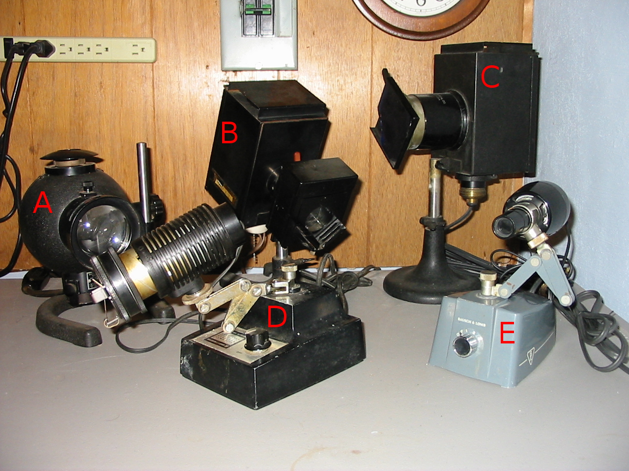
Bausch & Lomb microscope illuminators of various types
In the above photograph may be seen a variety of microscope illuminators. Each of the illuminators is capable of providing ample light and operates on AC current. One may infer from the variety of designs that each was intended to serve particular needs. Of those pictured some will serve for both transmitted and incident lighting∗, Köhler or critical illumination, microprojection, visual microscopy or photomicrography. Some are equipped with transformers to modify light intensity while others rely on filter holders of different sorts.
Illuminator A is a very high intensity lamp that is well suited for microprojection. It is equipped with a 200watt incandescent bulb which even with the light-baffled chimney and large airspace around the bulb gets quite hot after a sort time. Within the lamp housing a primary reflector is mounted behind the bulb and a two lens, friction fit, condenser system is mounted before it. The lenses of the condenser are fitted in a permanent relationship with each other and slide in or out of the body in unison allowing an image of the bulb, or a uniformly lit field to be focused at the required distance with ease. A slot in the rim of the condenser housing permits the use of various filters though only one may be used at a time. The stand permits the lamp to be raised sufficiently high to permit its use as a source of incident lighting. Due to the intense light provided by this illuminator it should not be used for ordinary visual work, but it can provide ample light for the highest power dark-field plate and film photomicrography.
B is a variety of illuminator that was once considered essential to effective microscopical work. It is able to provide Köhler illumination and features a large ventilated lamp housing with a baffle to keep excess light from the work-room. It is fitted with a standard medium base light bulb socket that may be adjusted to center the bulb or fillament in relation to the condenser system. Before the bulb is mounted a spiral focusing multi-lens condenser that terminates in an iris diaphragm. There is additionally mounted (yet not in contact with the diaphragm) a metal box in which is held a glass water-cell. A removable three filter holder is mounted to the water cell box allowing the use of filters from 2 too 3 inches, either square or round in any combination. A water cell may be filled with water simply to lower the physical transmission of heat along the path of light when observing sensitive specimens, but it may also be filled with chemical solutions to provide particular lighting characteristics†. Once the bulb and condenser have been properly aligned and centered one need only assure that the illuminator is the proper distance from the microscopes mirror when setting up for Köhler illumination. Although not so bright as illuminator A, this apparatus may be used for visual and photomicrographic work of any sort.
Next is seen illuminator C which is an earlier and less well outfitted example of much the same sort. The housing and condenser arrangement is nearly identical although the lenses in use are of significantly larger diameter. It does not have an iris diaphragm or water-cell although such accesories were available at the time of its manufacture. A single filter holder for use with 3¼ inch square filters is mounted at the terminus of the condenser. Although this example can not provide Köhler illumination when used with a powerful 100watt incandescent bulb it can provide critical illumination with sufficient light for photomicrographic, microprojection, and dark-field work. Like the previous example it is well suited for visual work.
D may be seen as a more modern and less complex version of B. It uses a low voltage high intensity halogen bulb mounted in a heat dissipating lamp housing on the end of an articulated arm attached to a heavy base providing adjustable power. The two lens condenser is spirally adjustable and carries an iris diaphragm and friction fit filter holder that permits the use of any combination of two circular or square 2 inch filters. This illuminator can provide high power Köhler illumination for all needs, excepting the most demanding microprojection work. Raised above the microscopes stage it can provide incident lighting for use with compound or dissecting microscopes. The only disadvantage of such an illuminator is that accompanying the use of a bulb that is not apt to be carried by local retailers.
Of those pictured the most commonly found is that designated E, a common Nicholas illuminator. The primary use of such a light source is in providing intense incident lighting for low power dissecting microscopes. Many varieties of microscope intended for low power work are fitted with holders in which the self contained lamp housing may be placed so as to provide a stable position that will remain constant from use to use. It is also common to use an articulated arm mounted to the transformer to allow positioning of the lamp as in this example. The rear of the lamp housing is designed to provide ventilation for the high intensity halogen bulb, while the narrow front portion bears two condensing lenses in a fixed relationship so as to provide a bright uniform spotlight with which to light an opaque object. An illuminator of this sort should not be used for visual work with transmitted light as even at the lowest power setting it is damagingly bright. A Nicholas illuminator will do nicely when using a long working distance objective on a compound microscope to examine an opaque object, the bright light can penetrate even strongly textured objects with ease.
Explanations
One will note that without exception all of the above illuminators are equipped with one form or another of condensing system. The use of a series of lenses integral to the illuminator eliminates the need for a bulls-eye lens between light source and microscope mirror as was common practice before the adoption of standard illumination methods. It additionally greatly reduces the time one will spend in setting up for critical illumination as only a limited range of focus will be possible so that one need not set the apparatus a different distance from the microscope from session to session. If practical it can be a great asset to fix both illuminator and microscope to a suitably large board the maintains them in the proper relationship.
All of the above illuminators may also be set up for critical illumination (A and E included, though it would certainly damage the users eye) so that an image of the lamps filament is in sharp focus with the image plane of the specimen. In practice, this would require that each of the bulbs used be of the clear glass variety. B and C are both fitted for use with a common medium base bulb which permits the use of bulbs of the sort one would use in indoor lighting. If one elects to use a standard lighting bulb a variety of different effects become possible. An opal white bulb may be used to obscure the image of the lamps filament and provide a more uniformly lit field of view. Specialty photographic bulbs‡ may be used to provide for the proper white balance with various photographic films. If however, one uses a frosted white bulb, it will be found that an image of the filament shows through, and all that will be achieved is the introduction of grain. It will generally be advisable to use only clear bulbs and then obscure the filament with a frosted glass filter as required, rather than purchase a variety of bulbs and be obliged to change from one to the other after waiting for the bulb to cool.
Looking at any of the above one might immediately recognize that the only exotic component is in most cases the lamp house and iris diaphragm. It would then be a simple matter to improvise a system of illumination that works as well if only one is willing to sacrifice the compact form represented in the above. Take for example a standard ceramic lamp base mounted to a board, place over it a large tea-tin that has had a hole of an inch or two diameter cut in one face coincident with the bulb. Cut a few slits on the opposite side over the bulb for ventilation and the housing is taken care of. One need only then mount two short focus lenses (Plano-convex arranged with the convex surfaces facing being optimal) at their primary focus from each other and provide some method of aligning them with the hole in the tin to obtain a means of achieving critical illumination. Introduce a fixed diameter diaphragm of appropriate size, and one can approximate Köhler illumination at the cost of a few dollars and an afternoons effort.
Notes:
∗Transmitted light refers to light which is passed (transmitted) through the specimen while incident light refers to light which falls upon (may be considered incident to) the specimen.
†At one time a solution of copper sulfate with various additives was used as the most accessible means of obtaining monochromatic light visually comparable with daylight.
‡For whatever reason color corrected bulbs for photography are less expensive that appropriately sized corrective filters.
Next time we will set up for Köhler illumination with a representative illuminator (B) and critical illumination with an other (C). -K
