I can not stress enough the importance of traditional micrography as a means of gaining understanding of the specimen, but for ooh and ahh factor digital is king. -K
The setup
For digital photomicrography a consumer grade mirror-less digital camera with removable lens is mounted over the eyepiece with a two part connector. The first part fits into the cameras lens socket and is friction fit to the second part which fits over the microscopes tube and rests upon its shoulder. The camera may be removed at any time without difficulty and all apparatus is away from the ocular so that it is un-obstructed and may be changed. With the camera removed the microscope may be focused as normal without recourse to the cameras display. As the imaging sensor is at the microscopes eyepoint no secondary focusing is required when the camera is moved into place.
The microscope is that same common workhorse that has been featured in the previous entries. Lighting is provided by a single 60 watt cool-white incandescent bulb in a goose-neck desk lamp. A 5x Huygenian ocular is used for every image. The camera is an older Nikon 1 J1. Anyone desiring to know the settings used for each exposure is advised to check EXIF data for each image. A traditional test object, the proboscis of a blow fly, is used for each image. Be warned, clicking on the images will display a full size (3872×2592) image of several megabytes size.
Photomicrographs
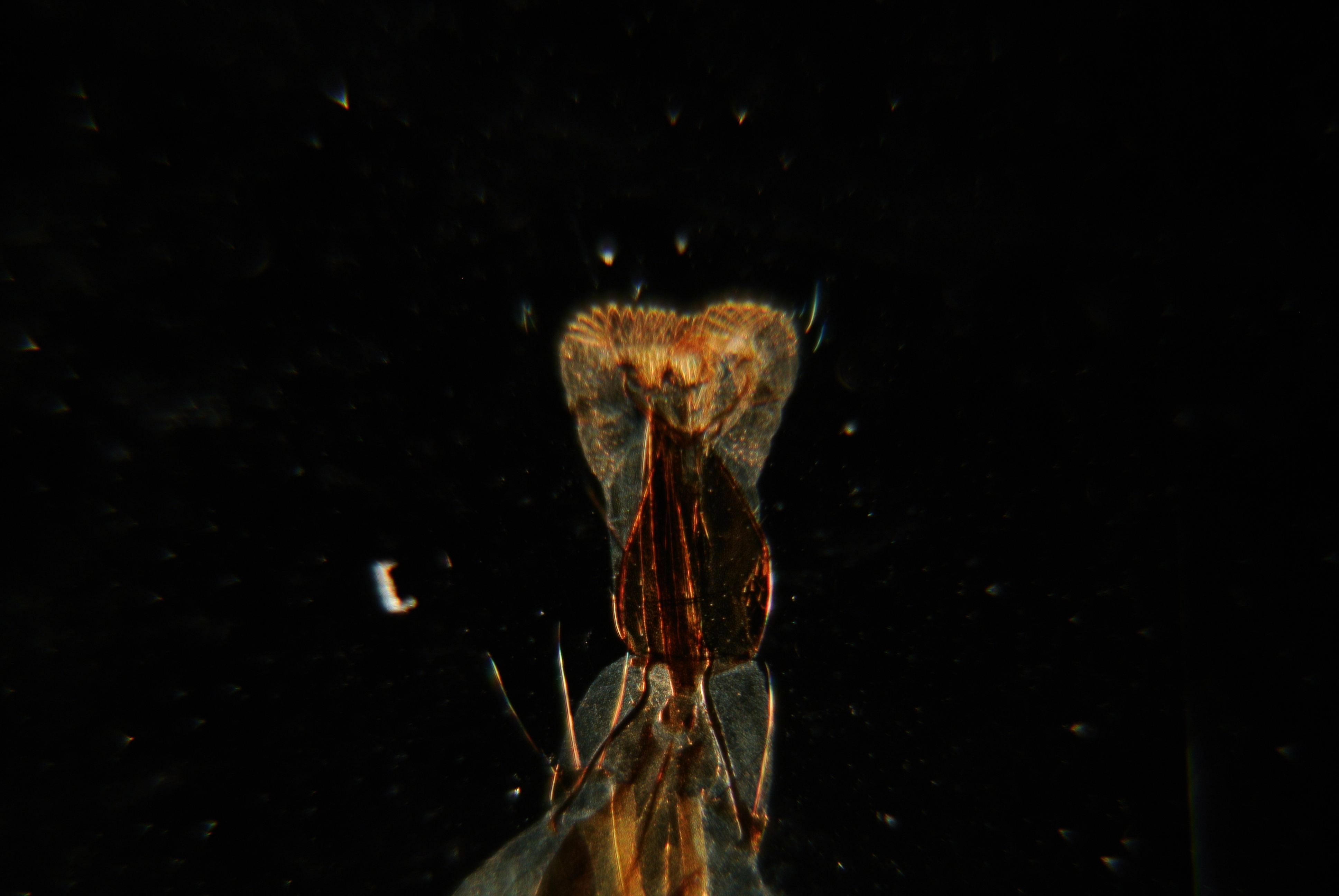
First we remove the lower portion of the divisible 10x objective leaving a perfectly serviceable 32mm equivalent focus objective. A larger aperture opening is spun into place with the circular diaphragm to avoid vignetting the image.
 Not bad considering the aberration inherent in such a lens. Observe that despite being relatively close to the center of the field of view the finer points of tung are out of focus.
***
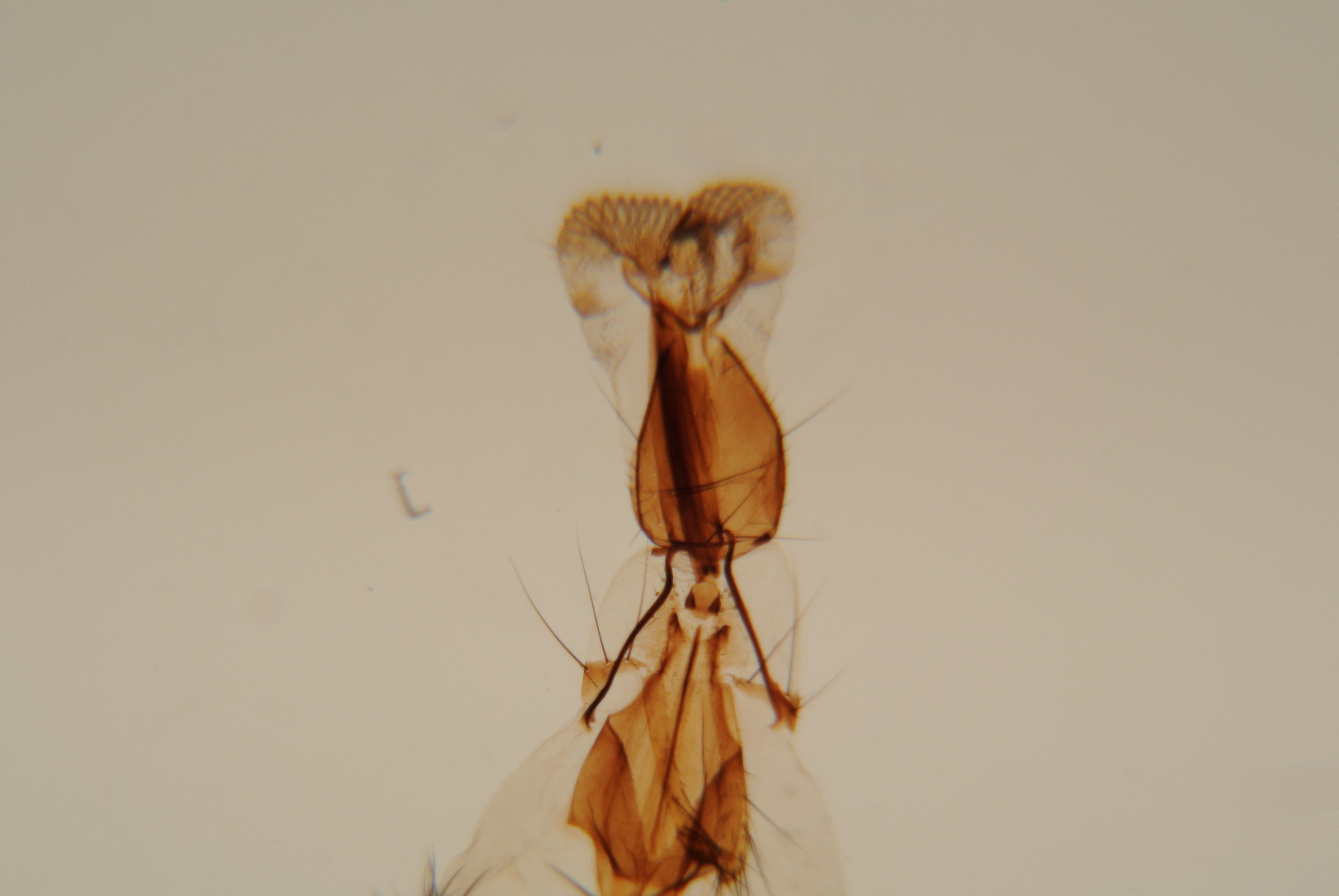
With the assembled 10x divisible objective (16mm EF 0.25NA) spherical aberration in particular is much less obvious. A smaller opening of the circular diaphragm is selected to provide better contrast and reduce glare. No realignment of the concave mirror was made.
 Much more of the structure of the tung may be made out although the depth of field is noticeably lacking. Only the smallest evidence of chromatic aberration is visible. Despite what is a very rudimentary lighting system the field is bright and even.
***
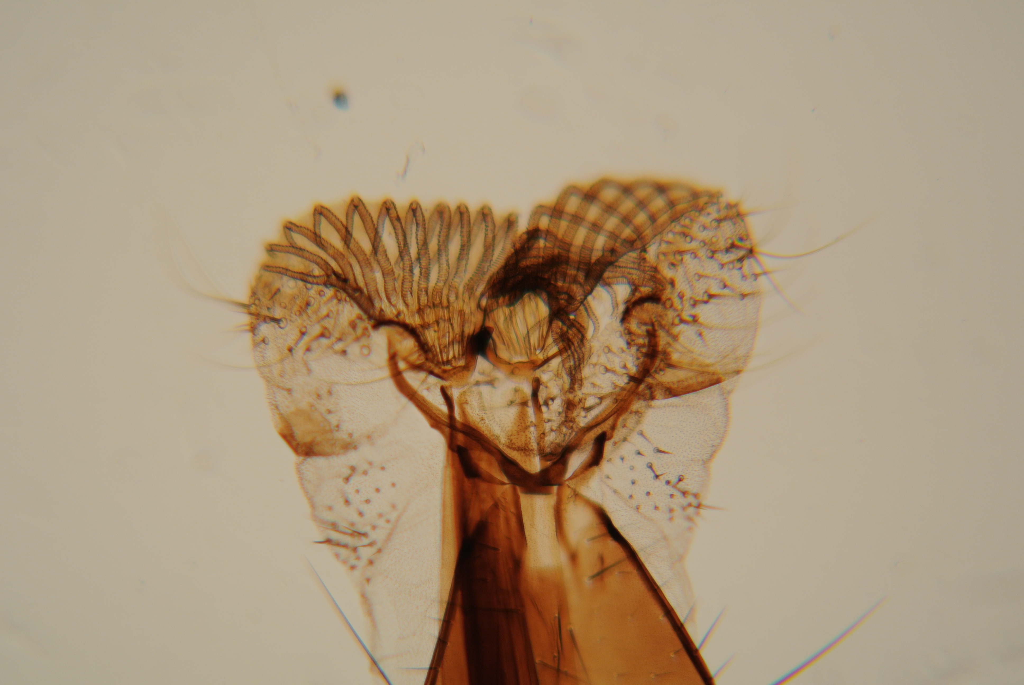
Switching to the 43x (4mm EF 0.65NA) objective we switch also to the smallest available aperture in our circular diaphragm. Unlike the previous change in magnification using the divisible objective the change from 10x to 43x is relatively parfocal and a few turn of the fine focus results in the following image.
 The field is surprisingly well lit for the absence of a condenser. One may observe that depth of field is more than one might expect especially when considering the relative thickness of the specimen. The color fringes of chromatic aberration are in evidence and anyone accustomed or intending to do much work at this magnification would surely be unsatisfied with the image. Understandably, one with experience might forget that those relatively new to the pursuit are less sensitive to such things and would likely be rather happy with the quality of the above image.
***
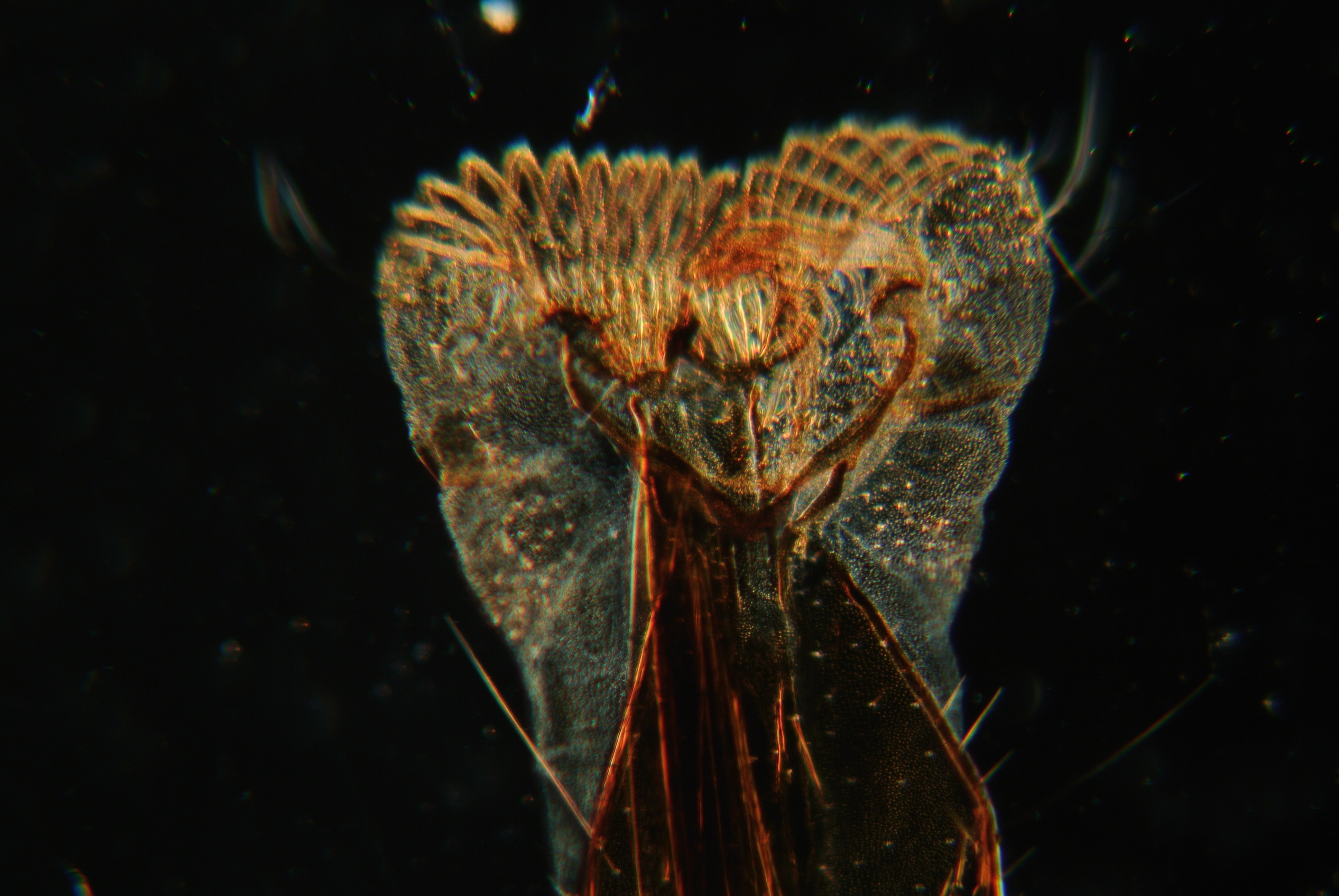
Turning the arm of the mirror sharply to one side and switching to the largest opening of the circular diaphragm we are able to take advantage of an all but forgotten lighting technique. A shade is placed so that the area below the diaphragm opening rests in shadow and the lower portion of the divisible 10x objective is removed.
 Oblique lighting is not generally possible with a substage condenser. Specialized stops may be put into place but even then the obliqueness of the light is subject certain limitations relating to the working distance and numerical aperture of the condenser. Here a starkly black background is visible because the surface of the table is black and out of focus for the objective. The specimen is brightly lit by the mirror and that light which it sends into the objective. Every speck of dust on the slide is noticeable and in the full size image one has no trouble at all in identifying to which side the mirror was swung (to the left).
***
With the fully assembled 10x objective a passable image is produced which is serviceable as a “poor mans dark-field”. Patch stops intended to render a standard condenser a dark-field substitute do not provide so dark a background.